Essentials of Embryo Transplantation in Liaoning Cashmere Goats
The Liaoning cashmere goat is originally from Gaizhou City. It is also known as Gaizhou Cashmere Goat. It has high cashmere yield, high net cashmere, long velvet fiber, moderate thickness, large body size, stable genetic properties, and improved low-yield goats. With its characteristics, its cashmere production ranks first in the country and it is known as the “National Treasureâ€. The application of embryo transfer technology to Liaoning Cashmere goats has greatly improved the breeding performance of elite ewes so that their superior traits can be fully utilized.
First, the choice of transplant time. The embryo transfer season is usually arranged during the estrous season of Liaoning Cashmere goats in order to maximize the potential reproductive performance of the sheep. In general, multiple choices are most suitable in the autumn of September-November.
Second, the surgical process of the recipient ewes
1, preoperative preparation: To prevent preoperative abdominal pressure is too large, resulting in surgical difficulties and the recipient's reproductive tract damage, 24 hours before surgery to stop feeding fodder, limited drinking water. Surgical instruments were soaked in a 0.5% benzbromide solution for 30 minutes prior to use. Supine Baoding recipient ewes. General anesthesia was then performed by intramuscular injection of 0.03-0.05 ml/kg body weight with 2% Jingsongling.
2. Surgical site treatment: The surgical site was selected approximately 2 cm from the anterior end of the breast. Both sides of the abdominal midline and the abdominal midline can be surgically sheared on both sides. The range is approximately 10 cm and 20 cm. Wash the area with soapy water or 0.1% benzalkonium chloride. At the same time shave the frizz with a scalpel and then clean it with clean water. First use 2-4% iodine to disinfect the surgical part from the inside out, and deodorize with alcohol cotton, then cover with a towel to prepare for surgery.
3. Surgical opening: Parallel to the midline of the abdomen, a 6-8 cm long incision was made in the longitudinal direction of the surgeon with a scalpel. After the skin is incised, the subcutaneous connective tissue is separated, the sarcolemma is exposed, the sarcolemmal is incised with a scalpel, the muscle layer is exposed, and the muscle layer and the peritoneum are bluntly peeled with the back end of the handle, and the middle finger and the index finger are separated. Into the pelvic cavity in front of the bladder touch the uterine horn, gently pull the uterine horn and ovary out of the incision, and plug the wound with two pieces of hemostatic gauze, wet the gauze with saline, to fix the uterine horn and ovaries so that it can not Retract the abdominal cavity.
Third, draw embryos. Use a transplanter to first aspirate a 1 cm long preservation solution, then take a 0.5 cm piece of air, and then take up the embryo. The liquid column containing the embryo should not exceed 1.5 cm.
IV. Embryo transfer
1. Fallopian tube transplantation: Peel the tubal umbrella on the side of the corpus luteum, find the orifice, and insert the transplanter with the embryo from 3 to 4 cm. Then gently push the embryo into the fallopian tube.
2, uterine transplantation: the uterine horn with a side of the corpus luteum is taken out, a 16-point needle with a blunt tip is punched in the uterus wall, the implanter with the embryo is inserted into the uterus from this hole, and extends to the tip of the uterine horn. Push the embryo gently. Be careful not to stick the implanter into the uterine wall. Whether it is a fallopian tube graft or a uterus graft, the amount of fluid should be limited to 20 microliters. Immediately after the transplant, check whether there are embryos left in the transplanter and confirm that there are no leftovers.
Receptor wound suture. After transplantation, the viscera was sprayed with a certain concentration of heparin solution and the clot was washed off. The viscera was rinsed with 25-30°C physiological saline and 250 ml of metronidazole (25-30°C) was injected into the abdominal cavity to prevent adhesions. Three layers of sutures were used for recipient ewes wounds. In other words, the peritoneum and muscle layers were sutured consecutively and the skin nodules were sutured. The interval between the needles was 1 cm. When stitching, sprinkle a proper amount of penicillin and streptomycin powder between the muscle and the skin to prevent wound infection.
Sixth, postoperative management. Ewes need to be observed after 24 hours to grazing with the herd. Postoperative 3-5 days, twice daily penicillin 800 000 units / time, streptomycin 1 million units / time, to prevent postoperative infection. In case of infection, care is given according to general surgical methods.
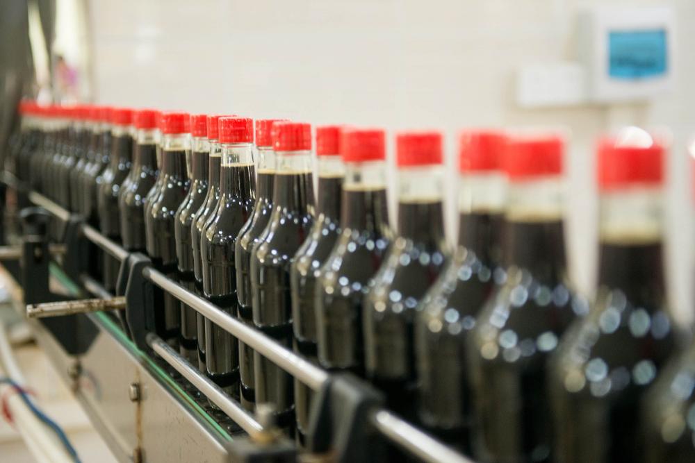
Lishida Superior Soy Sauce,as a popular condiment for cooking,is brewed from non-GMO soybeans and other high quality ingredients.We adhere to brew naturally and Chinese traditionally for 20 years,and all of our products are packaged by advanced production line.Lishida Superior Soy Sauce Series includes Light Soy Sauce,Dark Soy Sauce,Mushroom Dark Soy Sauce,Steamed Fish Soy Sauce,Gluten-Free Light Soy Sauce,Gluten-Free Dark Soy Sauce,Less Salt Light Soy Sauce,Less Salt Dark Soy Sauce and so on.

Not only passing the Quality Standard Certificate,we also have been awarded ISO9001 Quality Management System Certificate,HACCP System Certificate and so on,maintaining strict quality assurance.


Share the fresh with our love.We do hope everyone could enjoy fantastic moment with cuisines.
Superior Dark Soy Sauce,non GMO Soy Sauce,Superior Light Soy Sauce,Gluten Free Soy Sauce,Less Salt Soy Sauce
KAIPING CITY LISHIDA FLAVOURING&FOOD CO.,LTD , https://www.lishidafood.com